Assessment of infectious keratitis

Note: Infectious keratitis is a sight-threatening emergency. Urgent referral to an ophthalmologist is essential.
Infectious keratitis is a sight-threatening condition involving infection of the cornea. Causes include bacteria (eg Staphylococcus aureus, Streptococcus pneumoniae, Pseudomonas aeruginosa) and viruses such as herpes simplex virus (HSV) and varicella zoster virus (VZV) – see Comparative features and assessment of types of infectious keratitis for comparative features. Keratitis caused by fungi, mycobacteria or Acanthamoeba species is rarer and more difficult to treat.

Differentiation between types of keratitis is difficult and requires slit lamp examination, so prompt referral to an ophthalmologist for assessment is recommended.

Table 1. Comparative features and assessment of types of infectious keratitis

Nguyen, 2019The Royal Victorian Eye and Ear Hospital (RVEEH), 2024The Royal Victorian Eye and Ear Hospital (RVEEH), 2022The Royal Victorian Eye and Ear Hospital (RVEEH), 2022Watson, 2020

Printable table

Type of infectious keratitis

Presentation and risk factors

Investigations

Bacterial keratitis

Presentation:

  • painful eye
  • red eye
  • watery eye
  • mucous discharge
  • reduced or blurred vision
  • photophobia
  • white spot usually seen on cornea

Risk factors:

  • contact lens wear
  • eye trauma
  • compromised ocular surface (eg following eye trauma)

prompt ophthalmology referral for consideration of a corneal scrape for culture and susceptibility testing

for contact lens wearers, culture of swab samples from contact lenses and cases

Herpes simplex keratitis

Presentation:

  • painful eye
  • red eye
  • watery eye
  • reduced or blurred vision
  • photophobia
  • itching
  • epithelial keratitis (dendritic ulcer)
  • geographical ulcer
  • stromal keratitis (stromal opacity, stromal ulceration, scarring)
  • endothelial keratitis (stromal oedema, keratic precipitates)

Risk factors:

  • previous HSV infection
  • active cold sores

prompt ophthalmology referral

fluorescein staining of the cornea to facilitate diagnosis of epithelial keratitis – see Dendritic ulcer caused by herpes simplex virus

antigen detection or NAAT (eg PCR) of a conjunctival swab

consider optometry services to aid in diagnosis if ophthalmology review is likely to be delayed

Herpes zoster ophthalmicus

Presentation:

  • painful eye
  • red eye
  • watery eye
  • reduced or blurred vision
  • photophobia
  • can also cause epithelial keratitis and stromal keratitis

Risk factors:

  • shingles (herpes zoster), especially if rash involves tip of the nose (Hutchinson’s sign)

ophthalmology referral

diagnosis is usually clinical

if diagnosis uncertain, swab inflamed conjunctiva for NAAT (eg PCR)

Note:

HSV = herpes simplex virus; NAAT = nucleic acid amplification testing; PCR = polymerase chain reaction

Figure 1. Dendritic ulcer caused by herpes simplex virus

Note:

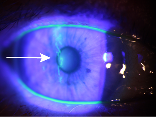
Photo of fluorescein staining showing a dendritic ulcer caused by herpes simplex virus.

Photo sourced with permission from Dr Alex Hamilton.