Erythema nodosum
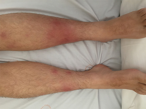
Erythema nodosum is a cutaneous reaction pattern occurring in the deep dermal and subcutaneous tissues. Lesions present as painful erythematous indurated plaques and nodules, usually on the lower legs (particularly the anterior tibial region); however, lesions can occur elsewhere. Patients may have fever, malaise and arthralgia. For a photo of erythema nodosum, see here. The lesions typically resolve over 2 to 3 weeks, leaving discolouration but no scarring. Less inflammatory chronic forms, particularly on the lower legs, may take months to resolve. The condition commonly recurs.
Erythema nodosum is associated with a range of conditions—see main causes listed in Main causes of erythema nodosum. The mechanism for the reaction is thought to be a delayed hypersensitivity reaction to various antigens.
|
Cause |
Examples |
|
bacterial infections |
beta-haemolytic Streptococci (especially following pharyngitis) (eg Streptococcus pyogenes, group C and G streptococcus), Mycobacterium tuberculosis, Yersinia spp., Mycoplasma spp., lymphogranuloma venereum, Chlamydia spp. |
|
other infections |
coccidioidomycosis, histoplasmosis, dermatophytosis, viruses (including hepatitis B and C, HIV, EBV and herpes simplex virus) |
|
inflammatory bowel disease | |
|
sarcoidosis |
– |
|
– | |
|
drugs |
sulfonamides, oral contraceptives |
|
malignancies |
lymphoma, leukaemia |
|
pregnancy |
– |
|
idiopathic |
– |
|
Note: EBV = Epstein–Barr virus; HIV = human immunodeficiency virus
| |
The condition is usually diagnosed and managed by a specialist relevant to the associated systemic disease (eg dermatologist, rheumatologist, gastroenterologist, infectious diseases physician).
Diagnosis is usually clinical. A thorough history and examination is required to identify the cause. Reasonable diagnostic tests include a full blood count, erythrocyte sedimentation rate (ESR), standard biochemical analysis, throat swab, streptococcal serology and chest X-ray. Biopsy is generally not required; however, if a biopsy is performed, the sample needs to be deep and include fat. Biopsy of inflammatory lesions on the lower legs is often associated with scarring and slow healing.
First-line therapy for managing erythema nodosum is bed rest and nonsteroidal anti-inflammatory drugs (NSAIDs). Leg elevation, compression bandages and stockings may assist recovery, particularly in lower leg erythema nodosum. The cause (eg tonsillitis) should be treated, if possible.
If symptoms are severe and the patient has no contraindications to oral corticosteroids (eg related to the cause), consider:
prednisolone (or prednisone) 25 mg (child: 0.5 mg/kg up to 25 mg) orally, once daily for 2 weeks, then taper the dose according to response. prednisolone prednisolone prednisolone
If there is any doubt about diagnosis, seek specialist advice.