Presentation of tinea
Tinea (ringworm) is caused by dermatophytes, which can infect the skin, scalp or nails (for treatment of tinea of the nails, see onychomycosis).
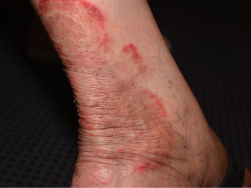
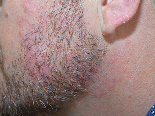
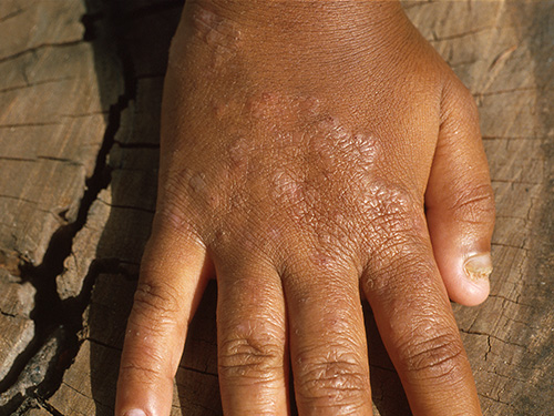
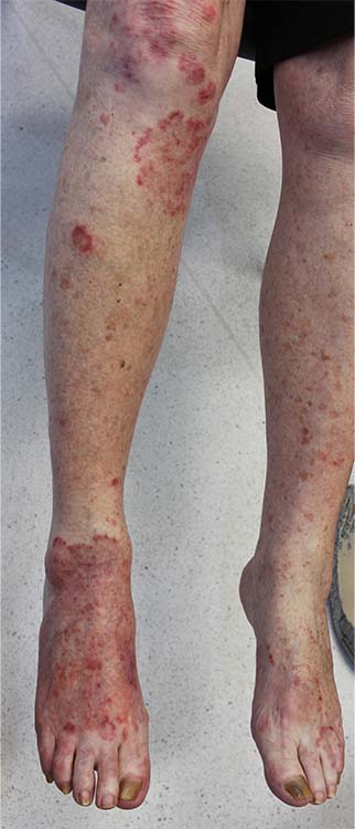
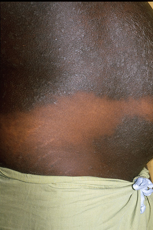
The typical rash is annular or arcuate (with a definite edge and central clearing as it expands), scaly and itchy. See here for photos of tinea. The more inflammatory and acute presentations of tinea are usually caused by animal species of dermatophyte. Tinea can be difficult to distinguish from dermatitis and other annular or patchy conditions (eg pityriasis rosea, granuloma annulare, psoriasis).
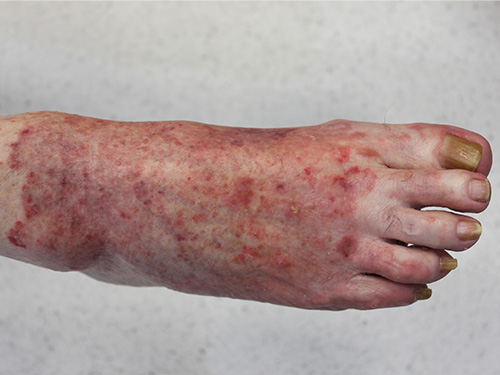
Tinea of the feet (tinea pedis) is particularly common in adults, but less so in children—it is often associated with tinea of the groin (tinea cruris). Tinea pedis can present as maceration in the lateral toe webs, as localised small blisters on the feet, or as diffuse scaling on the soles. Infection often spreads to the toenails, which become discoloured, thickened and dystrophic.
Tinea of the scalp (tinea capitis) mainly occurs in children. A kerion is an acute form of tinea capitis, presenting as a boggy, painful, inflammatory pustular mass with associated alopecia. For photos of tinea capitis and kerion, see here.