Presentation of melanoma
Cancer Council Australia Melanoma Guidelines Working Party, 2020Swetter, 2019
Melanomas are malignant tumours derived from melanocytes, usually on the skin. Occasionally, a primary melanoma develops in other organs that have melanocytes (eg eyes, genitals, oral mucosa, bowel). Melanoma most commonly occurs in sun-exposed areas, such as the nose, cheek, ears and upper backWee, 2020.
Australia has the highest incidence of melanoma in the world. Melanomas are a major cause of morbidity and mortality in young adults. The main risk factors for melanoma are:
- multiple common melanocytic naevi
- dysplastic or atypical naevus syndrome
- family history of melanoma
- blistering sunburns as a child or adolescent
- history of melanoma or nonmelanoma skin cancer
- solarium use
- fair complexion and a tendency to sunburn
- marked sun exposure (eg during work and leisure time) and solar skin damage
- immunodeficiency.
The Melanoma Institute Australia website provides risk prediction tools and other useful resources to aid in clinical decision making and discussing melanoma with patients.
Melanomas are often identified as a new or changing lesion by the patient or their partner. Thorough skin cancer assessment with adequate lighting and magnification with a dermatoscope can help detect lesions at an early stage.
Warning signs that a lesion might be a melanoma are listed in Melanoma warning signs.
Consider melanoma when a lesion is:
- new or changing (most melanomas arise de novo rather than from a pre-existing mole)
- prominent and pigmented, and stands out from other moles (‘ugly duckling’)
- rapidly growing (applies to a nodule of any colour)
- of particular concern to the patient
- atypical on dermoscopy (eg asymmetric pigmentation, blue-white veil, multiple brown dots, pseudopods, radial streaming)
- changed on sequential dermoscopy.
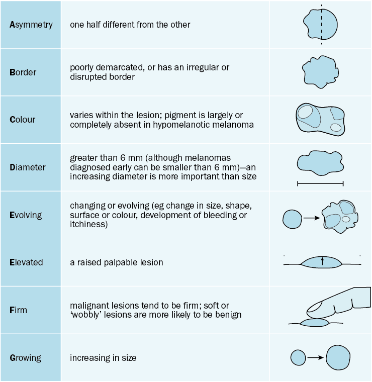
The ABCDEFG rule for melanoma diagnosis is a valuable tool, especially for patients—see The ABCDEFG rule for melanoma diagnosis. A small but significant number of melanomas cannot be diagnosed clinically, and a history of change (evolution) in the lesion can be the only clue.
Once a patient is diagnosed with melanoma, they are staged according to the features of the primary tumour and any regional lymph nodes. Printable PDFs detailing melanoma staging are available from the Melanoma Institute Australia website.