Types of melanoma
Lentigo maligna typically occurs on severely sun-damaged skin. It mainly affects older patients, but occasionally it occurs in adults younger than 40 years. Lentigo maligna is an in situ melanoma (confined to the epidermis), and there is often a long interval before it becomes invasive. Patients are often aware of an irregular, brown-to-black facial macule or patch for many years, so lentigo maligna can be quite large at presentation, even though it is still in situ. Histology distinguishes it from other pigmented facial lesions (eg solar lentigo, flat seborrhoeic keratosis) and identifies any invasive component (lentigo maligna melanoma). Dermoscopically, lentigo maligna may have an annular-granular pattern, asymmetric pigmented follicular openings and rhomboidal structures. Invasive melanoma can develop unpredictably, so active treatment of lentigo maligna is recommended. See Lentigo maligna for a photo of lentigo maligna.
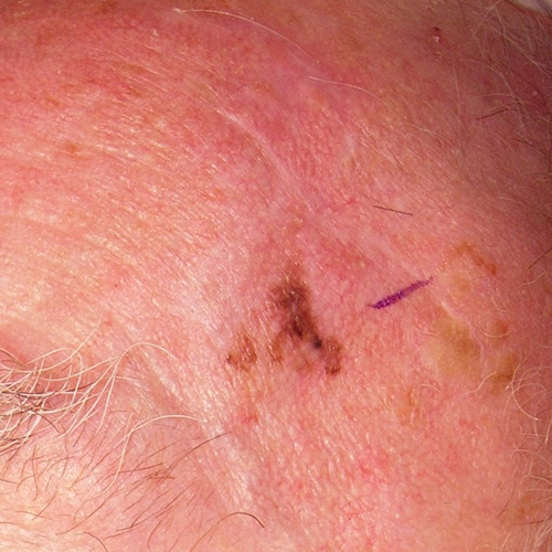
Photo sourced with permission from Dr Michelle Goh.
Reproduced with permission from the A-Z of Skin [digital]. Australasian College of Dermatologists. Sydney. https://www.dermcoll.edu.au/
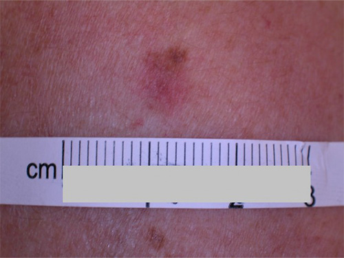
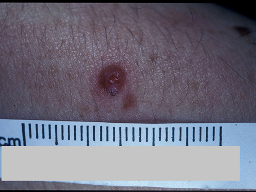
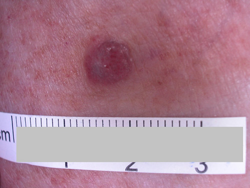
Superficial spreading melanoma is the most common type of melanoma, usually presenting as an irregularly pigmented macule or plaque. Dermoscopically, there is typically a range of colours, with architectural disorder, radial streaming, branched streaks, pseudopods, peripheral black dots and blue-white structures. See Superficial spreading melanoma for photos of superficial spreading melanoma.
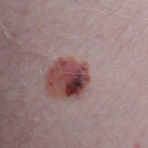
Photo sourced with permission from Dr Michelle Goh.
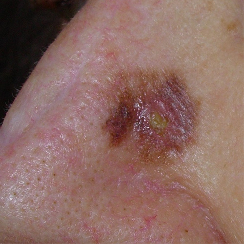
Photo sourced with permission from Dr Michelle Goh.
A nodular melanoma is aggressive and invasive, and can grow rapidly over weeks and ulcerate. The colour varies from black through to red. Approximately 50% are hypomelanotic. A nodular melanoma often defies the ABCDEFG rule, in that only the EFG (ie ‘elevated, firm, growing’) signs are appropriate. Rarely, a nodular melanoma is pedunculated and can resemble a pyogenic granuloma or basal cell carcinoma. Dermoscopy is less useful for a thick nodular melanoma. See Nodular melanoma for photos of nodular melanoma.
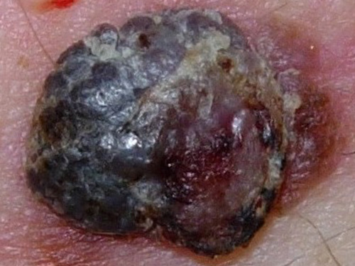
Photo sourced with permission from Dr Michelle Goh.
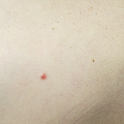
Amelanotic nodular melanoma. Photo sourced with permission from Dr Michelle Goh.
Reproduced with permission from the A-Z of Skin [digital]. Australasian College of Dermatologists. Sydney. https://www.dermcoll.edu.au/
Reproduced with permission from the A-Z of Skin [digital]. Australasian College of Dermatologists. Sydney. https://www.dermcoll.edu.au/
Acral lentiginous melanoma is the most common type of melanoma in people with dark skin types. It presents on the palms, soles or nail bed. The unusual location of these melanomas can delay diagnosis; tumours are often already large when recognised. See Acral lentiginous melanoma for a photo of acral lentiginous melanoma.
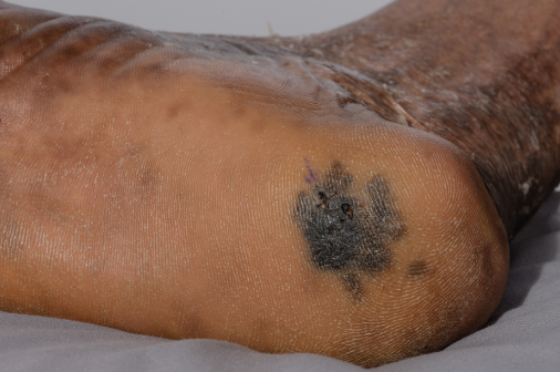
Photo sourced with permission from Dr Michelle Goh.
Desmoplastic melanoma is rare and aggressive. Its appearance is often subtle, and sometimes scar-like. Most are nonpigmented. Desmoplastic melanoma may be associated with overlying lentigo maligna.