Assessment of onychomycosis
Onychomycosis is a nail infection caused by dermatophytes, nondermatophyte moulds, and yeasts. It most commonly presents as tinea caused by dermatophytes (eg Trichophyton rubrum, Trichophyton mentagrophytes var. interdigitale, Microsporum); there may be more than one organism present on culture. Onychomycosis infections are frequently chronic and can be difficult to treat.
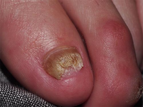
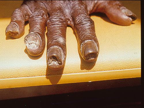
Onychomycosis caused by dermatophytes almost always presents in toenails, rather than fingernails. Clinical signs of onychomycosis include nail discolouration, hyperkeratosis and onycholysis. Initially, there is distal subungual onychomycosis (hyperkeratosis of the internal surface of the distal nail plate and of the distal nail bed [hyponychium]). Progressive nail involvement causes total dystrophic onychomycosis. Infection with T. mentagrophytes var. interdigitale may also cause an unusual form of onychomycosis called white superficial onychomycosis. See here for a photo of onychomycosis. Also see here for a photo of tinea affecting both the hand and nails.
Many nail disorders mimic onychomycosis (eg bacterial infection, psoriasis, dermatitis, lichen planus, viral warts, onycholysis, onychogryphosis); before starting treatment, confirm the diagnosis and causative organism with microscopy and culture. Take an adequate sample of the affected nail—ideally use a combination of clippings (of the distal nail plate) and scrapings (from under the nail for thickened crumbling nails, or from the nail surface). For a video of correct technique for obtaining nail specimens, see the Mycology Online website. A combination of microscopy and culture is used for diagnosis to reduce risk of false negative results. Consider the risk of sampling error from samples taken of areas without infection. Repeat samples may be needed.