Congenital melanocytic naevi
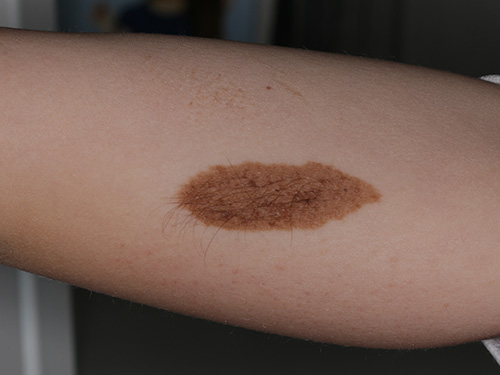
Melanocytic naevi (moles) are benign localised proliferations of melanocytes on the skin or mucosae (see Melanocytic naevi). Congenital melanocytic naevi are melanocytic naevi that are present at birth. It is a type of pigmented birthmark affecting approximately 1 in 100 children. Most congenital melanocytic naevi are initially flat or minimally raised at birth. Over time, they may become more lumpy, and can often grow hair. For a photo of congenital melanocytic naevi, see here.
Congenital melanocytic naevi are classified according to their projected adult size: small (less than 1.5 cm diameter), medium (1.5 to 20 cm diameter), large (20 to 40 cm diameter), and giant (more than 40 cm diameter)Krengel, 2013Nevus Outreach. Large and giant lesions are rare. To calculate projected adult sizeKrengel, 2013Nevus Outreach:
- if on the head: size at birth x 1.7
- if on the upper trunk or upper limbs: size at birth x 2.8
- if on the lower limbs: size at birth x 3.3.
The risk of melanoma in patients with small- and medium-sized congenital melanocytic naevi is low and considered to approach the same level of risk as for the general population. Prophylactic removal of the birthmark does not eliminate the risk of melanoma development in adulthoodKinsler, 2008Kinsler, 2017Krengel, 2006. Small and medium-sized congenital melanocytic naevi may be surgically excised if they are irritating or are a cosmetic concern—refer the patient for dermatologist advice. Laser therapy is not effective.
Large- and giant-sized congenital melanocytic naevi have been associated with an increased risk of melanoma development (10 to 15% lifetime risk), especially if they are accompanied by multiple smaller satellite naevi at birthKinsler, 2017. When melanoma develops, it usually occurs before the child is 10 years old. The melanoma may present as a deep nodule in the naevus or at extracutaneous sites (eg in the meninges).
The presence of multiple congenital melanocytic naevi (of any size) is also an independent risk factor for neurological complications (eg neuromelanosis, central nervous system tumours and malformations, hydrocephalus, seizures, abnormal neurodevelopment)Kinsler, 2008Kinsler, 2017Waelchli, 2015.
Refer infants with large or giant naevi, or multiple congenital melanocytic naevi (of any size) for paediatric dermatologist advice. Management is complex and multidisciplinary.