Examining the ulcer or wound
Assess and document the wound at the initial presentation and at each review. The wound characteristics (eg appearance, location) aid diagnosis. The status of the wound base, the amount of exudate, and the condition of the surrounding tissues influence the selection of appropriate therapies, including dressings.
Observing removal of the dressing provides useful information for assessing current therapy. Observe whether the dressing adheres to the wound, if there is pain on removal, and the quantity, appearance and odour of any exudate.
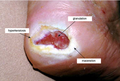
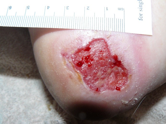
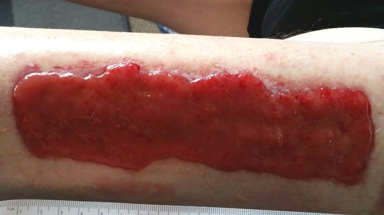
Note and record the wound’s location and dimensions (measure length, width, depth). Also note and record other important characteristics of the wound using the TIME acronym of tissue, infection/inflammation, moisture balance and edge (see Wound characteristics summarised using the TIME acronym). Wound charts, serial tracings and photographs are used to document wound characteristics. Photographs are also useful for communicating with other healthcare professionals; see Practice points for ulcer and wound photographs. Examination and documentation remains important at subsequent visits.
- Take photographs at the initial visit, then every month (more frequently if a change is identified), and on healing.
- Explain the process and use of photos to the patient and obtain consent.
- Cleanse the wound before taking a photograph.
- Ensure infection control procedures are maintained (eg take gloves off before using a camera).
- Use a white background to minimise shadows and glare.
- Ensure adequate lighting so wound details are clear—the flash may be required.
- Take the photograph from directly in front of the wound; holding the camera at an angle can distort the wound size and shape.
- Take a photo of the wound that includes a wide area of healthy tissue, so the location is clear.
- Then take a close-up photo to provide more detail.
- Ensure the wound measuring scale is visible in the picture (if it is causing significant glare, also take a close-up photo without the measuring scale).
- Include the date the photograph was taken (write the date on the measuring scale, or, if available, use the date stamp function of the camera).
- Take repeat photos from the same reference point as previously used.
- Always consider and maintain patient privacy. Do not include identifying patient features.
- Photographs must be stored appropriately and only shared professionally.
|
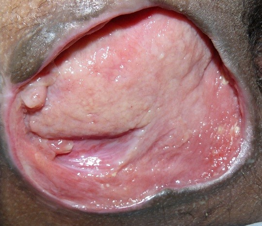
Tissue |
Tissue refers to the ulcer base. Note the presence and approximate extent of tissue types in the wound. This might include:
|
|
Infection/inflammation |
Determine if the wound is inflamed or infected. A superficial infection or increased bacterial burden in a wound can be indicated by the following features:
A deep infection can have any of the features above, and may include the following:
See also c_lwg2-c18-s2-1.html#lwg2-c18-s2-1__tlwg2-c18-tbl2 for more signs and symptoms of wound infection. Also consider noninfective causes of inflammation. Seek specialist diagnostic opinion if necessary. |
|
Moisture balance |
Moisture balance refers to the amount of exudate. Note the following features:
|
|
Edge and periwound area (skin within 4 cm of the wound edge) |
The edge of the wound informs the initial diagnosis and provides information about healing. Note whether the edge is:
|